

Description
Anatomical model of skin cancer
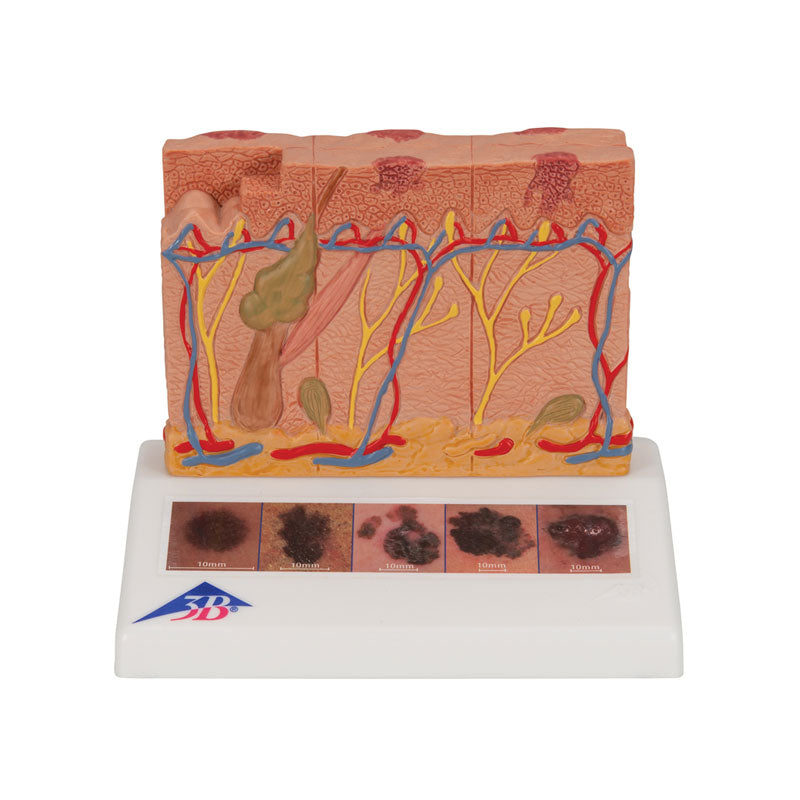
Anatomical model of skin cancer demonstrates six different stages of malignant melanoma. Seen from above, the model represents the visible skin lesions in the respective stages, making classification according to the “ABCDE” criteria possible.
Seen from the sides, the model represents the different depths of the melanomas, making it possible to classify according to Clark (levels I to V) and according to Breslow (measured in millimeters). The model holder contains 5 color illustrations, representing different forms and characteristics of malignant melanoma.
In this model, melanocytes:
• are found in the superficial layers of the epidermis;
• invade the entire epidermis, some proliferating into the papillary stratum;
• fill the papillary stratum;
• invade the reticular stratum;
• invade the subcutaneous tissue, while satellite cells are found near a vein.
Characteristics
- Weight: 0.34kg
- Dimensions: 14 x 10 x 11.5 cm
- Magnification: 8 times
Free Shipping
In purchases
amounts over €200*
Customer Support
By phone, chat
WhatsApp and Email
Secure payments
ATM, MB Way, VISA,
Paypal, Bank Transfer and Klarna.
Warranty and Returns
Transparent policy and
3-year warranty.